This morning, I walked into the lab, switched my sweater for a lab coat, then peeked into the microscope to check on my little blobs of human heart cells. They’re still beating! At least for now. Next week however, I’m going to give them heart attacks.
Scientists across the globe are trying to figure out how to repair and replace broken human hearts. As February is American Heart Month, I thought I would share a window into the front lines of heart research and cardiovascular regenerative medicine here in Winston-Salem.
 My name is Caleb Heathershaw, but my friends call me “The Heartbreaker”. I’m a second-year PhD student at the Wake Forest Institute for Regenerative Medicine. I work in the lab of Dr. Joshua Maxwell, spending most of my time building human cell-based in vitro models of heart attacks.
The Importance of Heart Research
Heart disease is the number one cause of death worldwide. For every five people that read this article, odds are, one of them will die from cardiovascular disease. That said, cardiovascular disease treatment is improving as new surgeries, procedures, drugs, and devices gain FDA approval and enter mainstream healthcare. Before treatments reach national impact, they have to be discovered, designed, and tested.
The heart is particularly challenging to treat, as it does not naturally regenerate.
Although axolotls and zebrafish can regenerate their hearts, our cardiac muscle cells (called cardiomyocytes) have limited regeneration. So, if those cardiomyocytes die, it is hard for them to grow back.
In the case of a heart attack, the coronary artery that feeds oxygenated blood to the heart muscle becomes blocked by a fatty plaque. This oxygen deprivation, or hypoxia, chokes out the cardiomyocytes, turning the tissue into a chunk of dead cells. Over time, stiff scar tissue forms and expands, making it quite hard for the heart to properly pump. My research is attempting to replicate the similar processes in a tiny lab dish.
Simulating a Heart Attack
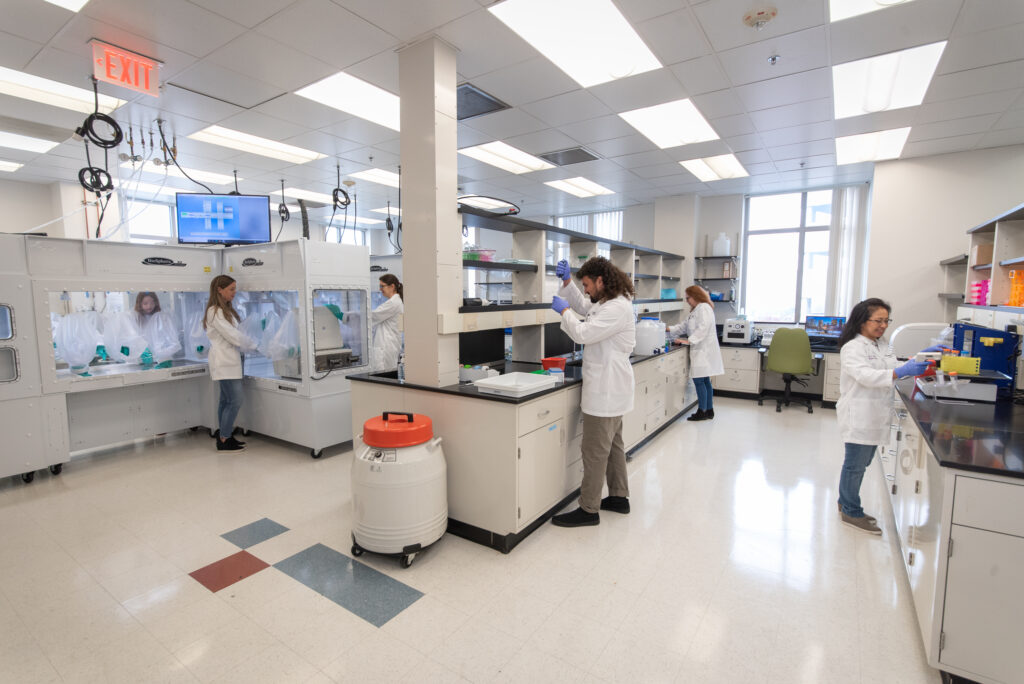
Next, we expose the heart organoids to hypoxia. This is done by moving the organoids into an incubation chamber filled with nitrogen and CO2 instead of oxygen. [For this procedure, I get to use an incubation chamber inside of the BioSpherix XVivo system in the RegeneratOR Test Bed. The Test Bed is a sandbox for scientists from across the community to test drive research tools as they’re being developed by biotech companies. It’s become an incredible asset for the regenerative medicine ecosystem.
WFIRM research associates, students and lab technicians keep the RegeneratOR Test Bed and its specialty equipment running smoothly.
Then, I assess the organoids’ health and see if they improve after different cell-based treatments. Different types of cells naturally send chemical and mechanical signals to other cells. So, if we can hunt down types of cells that send positive signals and harness those cells to release their signals in damaged areas of the heart, perhaps we can limit scarring and improve the health of cardiomyocytes that survived the initial heart attack.
My project isn’t the only one at WFIRM working on regenerative medicine approaches to treat heart disease. Dr. Maxwell’s team has also worked on using cardiac organoids as fibrosis models, atherosclerosis models, and genetic arrhythmia models. My lab neighbors in Dr. Sang Jin Lee’s lab are using bioprinters to create hydrogel cardiac patches that could be used to deliver cells directly to damaged tissue. Upstairs, Dr. Chris Porada’s group is even blasting heart organoids with space radiation to find ways of improving astronaut health.
The Future of Heart Regeneration
Most cardiovascular regenerative medicine research is still in the discovery stage. The treatments we are designing now will take years to perfect, prove, and produce before reaching patients. Good science takes time.
Two decades ago, heart organoids did not exist. Who knows where heart research will be two decades from now! Two decades ago, I was just a kid jumping around in the hills of Wilkesboro. Now, I’m a student scientist at one of the most exciting research institutes in the world, participating in the kind of slow, steady, deliberate heart research that changes things. So, I think I need to stop writing, roll up my sleeves, and jump back in the lab to tell my heart organoids to keep on beating, we’ve got work to do.
Happy Valentine’s Day and Happy Heart Month!
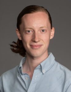
Author: Caleb Heathershaw
PhD Student at Wake Forest Institute for Regenerative Medicine